Definition of Euchromatin and Heterochromatin
Already during preparation under the stereo microscope it was possible to see that the polytene chromosomes were not equally condensed in all suspensor cells to the same degree. Even more pronounced these differences could be obvserved on base of the brightness in phase contrast microscope.
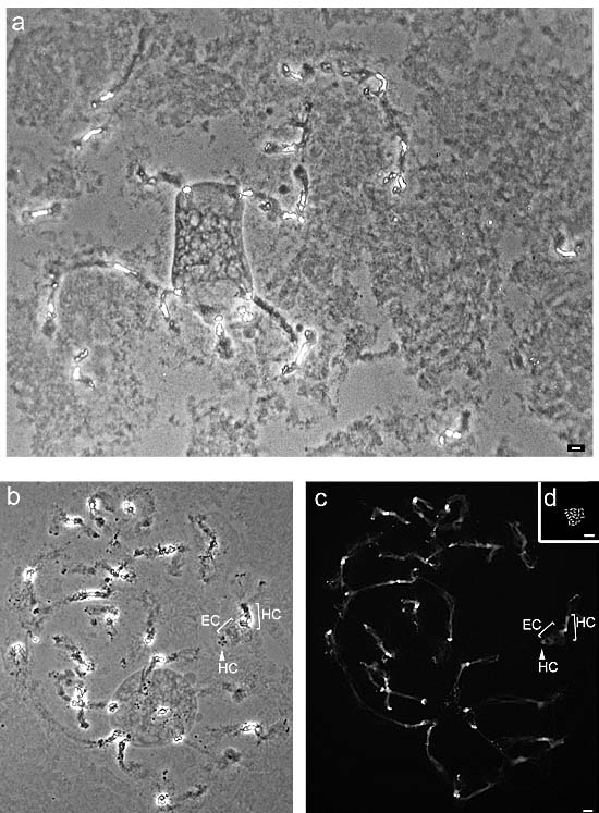
In approximately 90% of the nuclei chromosomes displayed segments of three different brightness levels. The centromeric region demonstrated mostly a particularly bright area whereas the chromosome ends displayed usually very dark spots and the intervening areas appeared in a medium gray, slightly darker than the Background (Figure a)
According to Heitz (1928), those areas are considered heterochromatin that remain
condensed even during interphase and that are stained more
intense compared to the mass of euchromatin. Applying the first part of
the definition to the above observation on polytene chromosomes of Phaseolus coccineus,
which represent interphase chromosomes, then the following
considerations can be concluded:
The highly refractive and therefore bright sections of the centromere region and
the dot-like structures at the end of the chromosome represent tightly package(condensed) chromatin and are hereinafter referred to as
heterochromatic.

The intermediate chromosome segments, that do not appear refractive and not dark represent loose or decondensed chromatin and can be considered therefor as euchromatic.
Comparing the intensity of brightness along the chromosomes of a cell nucleus between the phase contrast image and the after DAP fluorochrome staining, another concordance becomes evident. According to the above mentioned definition heterochromatic segment are stained more intense with DAPI than the euchromatic segments (see labled chromosome in figure b and c).
Previous studies showed that the morphology of the polytene chromosomes of Phaseolus coccineus can verify considerably, regarding both the total chromosome length as well as the distribution of heterochromatic bands in the euchromatin (Nagl 1965; Nagl 1967; Nagl 1981).
Therefore this study tried to find different features of the chromosomes which should enable a more reliable classification than before. Escpecially it should be possibile to identify each polytene chromosome after fluorescence insitu hybridization. For this reason the preparations for karyotyping undergo the same pretreatments with RNase and pepsin/HCl as the preparations for the fluorescence insitu hybridization and the same fluorochrome staining with DAPI and propidium iodide.

